Case Studies – Elsa
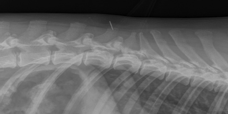
In February, Elsa appeared suddenly painful and restless. She was examined and mid-spine was found to be focal in her discomfort. She was given pain relief (buprenorphine and meloxicam) and advised to adhere to strict rest. The following day, she was experiencing ataxia and upon re-examination delayed hind-limb proprioception was identified. Radiographs showed spondylosis (Figure 1) throughout the lumbar region as well as moderate to severe bilateral hip dysplasia. It was advised rest and analgesia was to continue. Elsa was also prescribed gabapentin and previcox.
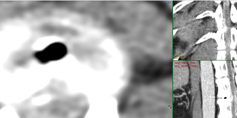
Elsa’s owner decided the next day, as her clinical signs were still worsening, to proceed with advanced imaging. One of our referral vets examined her again, identified grade II ambulatory paraparesis and slow pedal reflexes. A CT scan showed mild extradural cord compression at T12-13 (Figure 2) as well as multiple degenerative discs but none of these lesions causing cord compression.
A hemilaminectomy was performed wherein a large amount of disc material was removed. Fenestration of T12-13-disc space was performed to minimalise chance of further disc extrusion causing spinal cord compression.

Figure 1

Figure 2 Left: Axial view; the dark space depicts air which should not be there. This is a sign the disc extrusion has happened recently. Upper Right: The spinal cord (central, grey) is being compressed by disc contents (lighter colour). Lower RIght: sagittal view of air.
Elsa recovered very well, and remained hospitalised for ongoing analgesia, monitoring, rest and ambulation support. When she was more comfortable, passive physiotherapy was begun to encourage circulation and proprioception.
Within a week, Elsa was able to ambulate from sitting to standing without assistance and was actively moving her right hind limb. After another couple days she was taking steps and manging to walk with minimal support.
Elsa remained hospitalised for ongoing physiotherapy and within 2 weeks she was walking without support.
She was discharged with oral medications and a demonstration to the owner how to continue physiotherapy at home.
The following video was taken 7 days post-operatively:
The following video was taken 16 days post-operatively:
Physiotherapy session she was receiving at home:
After two weeks, her re-examination showed massive further improvements to her ambulation and proprioception. She was comfortable and her owner managing the recovery well. It was advised she returns to exercise over six weeks and hydrotherapy to be integrated into the rehabilitation plan.
Just under 8 weeks post-operatively, Elsa’s owner very kindly updated us on Elsa’s progress from a physiotherapy session she was receiving at home.
The progression in strength, range of motion and proprioception is huge. Elsa’s surgery resolved the spinal compression thanks to expert work by Kris, the surgeon, and her recovery was possible due to commitment from the nurses, complimentary therapists and of course, Elsa’s dedicated owner.


