Case Studies – Winston
Winston presented when his owners noticed a lump on his left-hand side. Winston was prescribed anti-inflammatories and imaging of his chest and abdomen was recommended. Soft tissue swelling was observed on radiographs. Fortunately, only one lesion was identified. It was noted that the mass was rapidly growing in the few days between first appointment and investigation. A surgical incisional biopsy was recommended and performed. Histopathology results were inconclusive, but
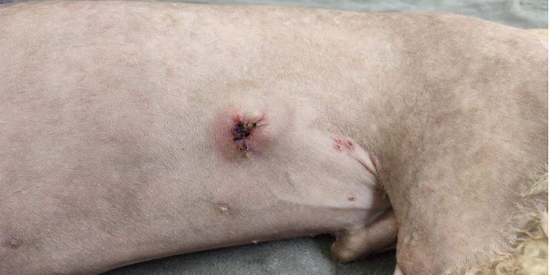
The mass after biopsies which confirmed subcutaneous haemangiosarcoma
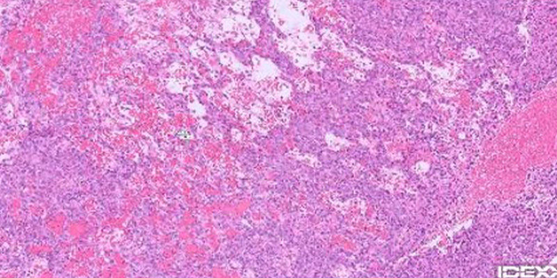
Histology slide showing invasive neoplasm – comprising dense streams of neoplastic cells rarely forming irregular blood-filled channels on a dense fibrovascular stroma
spindle cells were observed within the sample which caused concern this could be neoplasia. A further, deep tissue biopsy was necessary to confirm diagnosis. This was performed and urgent re-testing of the cells organised. The results showed highly malignant haemangiosarcoma. Without treatment, the risk of metastases is high and rapid mortality is almost certain. Both surgery and chemotherapy are recommended to treat and reduce occurrence of further tumours. It is important that a wide margin is included in the removal; or complete removal of malignant cells remain, reestablish and render the surgery ineffective.
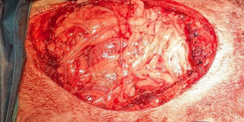
Surgical site wherein tumour removed with 3cm margin, full abdomen wall and 3 ribs were resected. Diaphragmatic transposition was also necessary
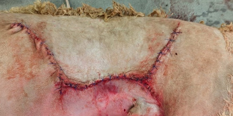
Advancement Flap used to close the defect
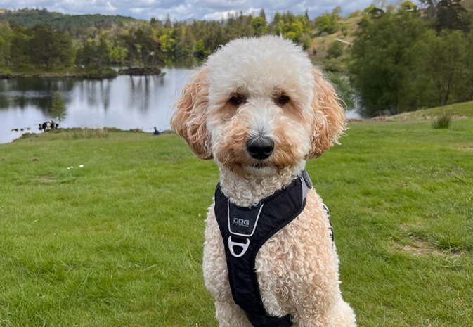
Post-operative histopathology showed the surgery was effective, likely extending Winston’s lifespan by at least 12 months. He also received a round of chemotherapy treatment following surgery. Winston returned to exercise 6 weeks after surgery and look at him now!


